For the full story, please see link:
http://www.sfweekly.com/2009-11-04/news/under-fire/
Exerpt from SFWeekly, "Under Fire," AnnaMcCarthy.
....Charles Lee, whom Estrada was visiting that day, was not one of those doctors. Lee, the director of microsurgery at St. Mary's, is a body reconstruction expert. He and his team received media attention in January 2008 when they successfully harvested a man's big toe to replace a thumb he had lost in a woodworking accident.
All expertise aside, Lee admits there were times he thought they would have to amputate Estrada's leg: "It was a pretty big wound," he said. "This is about as big as it gets." Because Estrada had lost so much muscle, the injury required a similar kind of tissue transplant as the toe-to-thumb surgery. When he first arrived at San Francisco General, his bones were sticking out of his uniform pants. Lee says he transplanted muscle from Estrada's abdomen to replace what he had lost on his leg; Estrada sports a long, dark centipede scar running down his belly to prove it.
Thanks to Lee and his crew, Estrada may have the opportunity to get back to work as a firefighter, which he says he wants to do — no matter how hard it is for him to watch the YouTube video of the fire, captured on a cellphone by a passerby. The video shows the entire incident from the moment Estrada approaches the warehouse with the hose to the moment he's loaded into the ambulance.....
This site is dedicated to educating patients about Reconstructive Plastic Surgery, its history, options, and relevance to Medicine and Surgery. Key words: Wounds, Trauma, Cancer, Breast Reconstruction, Infection, Osteomyelitis, Limb Salvage, Lymphedema, Hand Surgery, Microsurgery, Flaps, Skin Grafts, Negative Pressure
Friday, December 11, 2009
Sunday, May 31, 2009
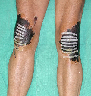
Frostbite after Postoperative Cryotherapy-- Wound Management & Limb Salvage


Picture #1: Skin Necrosis after Frostbite from Cryotherapy
Picture #2: After Debridement of Soft Tissues, use of Negative Pressure Dressing
Picture #3: Bilateral Free Flap (Rectus Muscles + Skin Graft) on both Knee Wounds
Picture #4: Bilateral Limb Salvage, Patient is Weight Bearing and Walking
Severe Frostbite of the Knees after Cryotherapy (Excerpts from original article by Lee CK, Pardun J, et al, Orthopedics 30(1):63-4, 2007 Jan)
"Abstract:
We present a case report of a patient who sustained full thickness soft tissue injuries over the bilateral knees after patellar tendon repairs and postoperative cryotherapy. The injury was severe enough to require bilateral microvascular free tissue transplants to cover both knee surfaces. Complications from cryotherapy have been reported in the literature, but are not common; this represents an extreme example. We review the literature and discuss treatment and prevention protocols.
Cryotherapy has been used to treat pain and inflammation since the
time of Hippocrates.1 Ice, snow,cold water, and cold compresses have been used to treat a multitude of soft-tissue traumas.2 More recently, cryotherapy
has been used increasingly in sports injuries and in the postoperative orthopedic setting.3 However, there have been a number of reports of complications from cryotherapy– most commonly frostbite and peripheral nerve injury–that point out itsbenefi ts but also its dangers.1-5 These previously reported complications has been diverse in location and severity. This article reports a significant complication of cryotherapy as a result of a relatively common regimen of application of ice packs to knees in a postoperative setting...
This case represents a severe frostbite injury after cryotherapy. With proper instruction and use of the cooling device, these complications are mainly avoidable. The patient had minimal padding between the cooling wrap and skin. In addition the patient used the device continuously for two weeks. It was likely that thermal injury occurred the moment the dressing was applied until the patient first took off the dressing two weeks later.
Frostbite occurs by the formation of ice crystals in the intracellular and extracellular space. During the cooling process, the extracellular ice crystals form and osmotic pressure increases, drawing water out of the cells. This leads to intracellular dehydration with an increase in intracellular electrolytes, proteins and enzymes which lead to cell death. Additionally, there is vascular endothelial damage leading to intravascular thrombosis and reduced blood flow. AV shunting occurs at the capillary level and end organ tissue damage is compounded. During the warming process, there is an influx of fluid back into the cells causing intracellular swelling. The warming process also allows reflow, vasodilation and reactive hyperemia to occur leading to increased inflammatory mediators, causing further cell death.
Cryotherapy works by three main processes. First is the reduction in the inflammatory process by inducing a hypometabolic state. Decreasing inflammation decreases the amount of cellular damage by inflammatory mediators, ultimately reducing the amount of capillary permeability and thereby decreasing edema. Second is the decrease in hematoma formation which is produced from vaso-capillary constriction and decreased blood flow. Finally is the induction of analgesia by cold. This is thought to be due to decreased nerve conduction velocity and decreased muscle spasm. In combination, cryotherapy is an ideal postoperative therapy which decreases pain, inflammation, hematoma, and the amount of postoperative narcotic usage...
Current recommendations for cryotherapy include 20-30 minutes of cryotherapy with a maximum time of 40 minutes, always with a protective covering (usually a towel) between the cryotherapy wrap and the skin. The cycle can be repeated every 2 hours while the patient is awake....
Although the incidence of complications from cryotherapy appear rare, estimated at 0.00225%,5 it is likely that this number is an underestimation as there are many unreported cases in conjunction with the increased use of continuous cryotherapy in the postoperative setting. We see this as an opportunity to emphasize the importance of education between the patient and doctor about this device and point out its potentially devastating risks.
Friday, May 29, 2009
Outpatient Wound Care Center in San Francisco 2009


San Francisco Wound Care & Reconstructive Surgery Center
We are proud to annouce the opening of an outpatient wound care center in San Francisco on June 1, 2009. It will consist of a multidisciplinary team of reconstructive plastic surgeons, orthopedic surgeons, general and vascular surgeons, podiatrists, endocrinologists and internal medicine, and dedicated wound nurse practitioners. Our team is composed of surgeons and physicians from the University of California, San Francisco (UCSF) as well as the community at St. Mary's Medical Center.
We will focus on all wound types, both acute and chronic, and of any complexity. These wounds can be from traumatic, vascular, infectious, metabolic, and cancer and radiation. We will have every tool and cutting edge option available to heal the simple to most difficult wounds.
Our office is at 450 Stanyan (Main Building of St.Mary's), 2nd Floor at the PROS Center. Telephone number is 415 750 5588. Email: sfwounds@gmail.com Please ask to speak to Josie Gomez, our wound nurse practioner.
Wednesday, April 1, 2009
Achilles Tendon Rupture, Wound, and Microvascular Tendon Reconstruction


Picture #1: Achilles Tendon Infection & Wound, 1 month after Rupture and Repair
Picture #2: Gracillis Muscle Free Flap as Vascularized Musculotendinous Reconstruction of Achilles Tendon Loss after Infection
Picture #3: Full Range of Motion of Ankle with Functional and Intact Achilles Tendon
Picture #4: Full Range of Motion of Ankle with Functional and Intact Achilles Tendon
The Achilles tendon is the largest tendon in the human body and a critical component in the function of the ankle joint to allow "push off" movement in walking and running. It is commonly ruptured during sports activities that require treatment by the orthopedic surgeon to repair the tendon. Usually the patient goes on to do well from this.
In some rare circumstances, the repaired tendon can become infected which can threaten the viability of the tendon. By the time we see the patient, the wound is quite large and severely infected. The patient is concerned about playing sports again.
Depending on the severity of the infection, multiple options are available to save this situation. Most importantly the wound needs to be explored and cleaned. Afterward the anatomy of the wound is further delineated. If the tendon loss is small, the wound up may be closed with local tissues and future tendon graft. If the tendon loss is moderate to large, a more complex reconstruction can be performed with both tendon and skin. This usually requires microsurgical expertise where tissue is transplanted to reconstruct the lost tissue.
We have used both the Gracillis muscle+tendon and the ALT (Antero Lateral Thigh) flap to reconstruct this complex defect with a high degree of success using microsurgical techniques. This requires an orthopedic and plastic surgery approach to combine their expertise to maximize functional recovery of the leg. Patients with Achilles' tendon ruptures are usually young and healthy men. Everything should be done, and all the options discussed to bring the injured leg back to as near normal function as possible.
Cutting Edge Treatment for Varicose Veins & Spider Veins

Painful, swollen legs with bulging "varicose" veins can be treated by several methods. In the past, vein ligation and vein stripping have been the standard. However, Vein Ligation has a high recurrence rate and Vein Stripping is an extremely painful procedure, often requiring an inpatient hospital stay, bed confinement for days, and long recovery.
Varicose veins can cause a variety of symptoms: Leg swelling, aching and pain, tiredness, skin changes, and even skin ulceration (venous stasis ulcers). These issues can lead to severe problems in the quality of life and activities of daily living, and in worst cases, severe infections which may threaten the viability or function of the leg.
Nonsurgical, EndoVenous Laser Treatment (EVLT) of Symptomatic Varicose Veins has become the treatment of choice with high success rates (> 95%), can be performed as an in-office procedure under local anesthesia, and patients walk out of the office immediately afterward. It is an extremely gratifying procedure.
As a Plastic Surgeon with expertise in Microvascular Surgery, Dr. Lee offers the highest level of technical precision to this safe, effective, treatment of Varicose Veins. Most often, these treatments are covered by Insurance.
www.sfveinlaser.com
Tuesday, February 24, 2009
What is an ALT flap?
The ALT flap is a very common, workhorse, flap used for various soft tissue reconstructionl. ALT stands for AnteroLateral Thigh flap. It is a skin flap that is harvested from the lateral thigh with the main blood vessels coming from the Deep Femoral Artery with a branch called the Lateral Circumflex Vessels.
Treatment of Chronic Wounds & Wound Care
There are many thousands of patients in the community with chronic wounds. The definition of a chronic wound is one that has not healed by six weeks time. As most of us know, wounds of all types eventually do close within a span of six weeks. This can range from small cuts and abrasions to larger open wounds of the legs or the abdomen from trauma or surgery. There are many patients in the community who have been undergoing local wound care for many months to years with minimal to no results. These wounds can degenerate into deeper infection such as osteomyelitis-- deep bone infection, and future loss of the limb.
Local wound care is very important to healing a wound. There have been many technological advances in newer dressings that have made wound care safer and more effective for the patient and health care provider. When a wound and has stopped healing in the six-week timespan, it is important to stop and reassess the wound. More than likely there are other causative factors that are preventing the wound from healing. This is when it is time to seek consultation from wound care experts.
As reconstructive plastic surgeons, we are the premier wound care experts. We have the ability to not only asses the wound, but have every option available to us to treat the wound. We are surgeons who deal with three dimensional anatomy. What this means is that we can assess the wound from a top-down and bottom-up approach that accounts for every part of the wound anatomy. Not all health professionals in wound care can have this approach as they cannot take the patient to the operating room to fully explore the wound and it cleanse the wound via débridement. The the ability to explore and debride a wound is the most critical factor in healing a chronic wound.
The options available to plastic surgeons include local wound care, negative-pressure (VAC, EZCare Negative Pressure, etc), to high-tech dressings such as Actcoat, Apligraf, Dermagraft, Integra, etc. After this we have the ability to place skin grafts that are split-thickness or full-thickness. We also have the ability to perform a local flaps which allow us to move tissue into the local area to close the wound. Finally, a plastic surgeon has the ultimate tool in reconstructive surgery--Microsurgery. We are able to transplant live tissue from another part of the body to the wound that requires this new tissue. We are able to reconnect the blood vessels to the transplanted tissues to close wounds that were never thought possible. Specifically, wounds that have been radiated or are chronically infected for many years have very few options for treatment. With microsurgical free tissue transfer, we have all of the options available in the reconstructive ladder to heal any wound, of any type, and size.
I encourage those who have been living with difficult wounds in their life to seek consultation with us to see if there are better options than the status quo of a nonhealing chronic wound.
Treatment of Osteomyelitis in the Extremity


Osteomyelitis is a severe and devastating bone infection. This can occur from simple wounds (venous stasis ulcers) to severe trauma of the extremity from open fractures.
In our practice, we find that many patients have not been adequately treated for osteomyelitis. In our view, osteomyelitis is a surgical disease. This means that IV antibiotics and other lesser therapies (such as hyperbaric oxygen) have only a minor role in the true treatment of this disease. The only sure way to eradicate osteomyelitis is to debride all necrotic and infected tissue, including the bone. The reason why this is not always done is because of the concern for removing bone , and soft tissue, which is not always replaceable.
At the PROS Center, we worked hand in hand with our orthopedic colleagues to have an OrthoPlastic approach to osteomyelitis. This means that we have our orthopedic surgeons, as the bone specialists, to remove all of infected bone. As plastic surgeons, we then have the expertise to provide all of the options for stable soft tissue coverage. Once the soft tissue envelope is stabilized, the orthopedic surgeons may return to perform other ancillary procedures to replace or stabilize the bone.
There are a few centers in the country, where there can be one-stop shopping to treat these extremely complex disorders such as osteomyelitis.
Subscribe to:
Posts (Atom)