Wednesday, June 22, 2011

Preventing Total Knee Arthroplasty Infection with Microsurgery -- The Prophylactic Free Flap

Total knee arthroplasty (TKA, knee replacement surgery) is a common procedure to treat knee pain and arthritis.  Over 120,000 knee replacement surgeries are performed per year in the United States.

The surgery involves making a midline incision over the knee joint and allowing the orthopedic surgeon access to the joint area to place an implant that acts like a joint.  Once the joint is in, the skin is  closed over and allowed to heal.  Often, the knee is started on a range of motion protocol to prevent stiffness.

On rare occasions, the knee skin may not be viable, sturdy, or durable to allow the surgeon to perform the knee replacement surgery because the closure of the soft tissue may be difficult   (history of trauma with scar, thin skin, skin grafts, psorasis, etc)  .  If the skin can be properly closed over the implant, this is a serious situation for the viability of the implant and the leg.  An exposed implant is an infection and can lead to serious complications.

At our center, we have worked with several of our orthopedic surgeons who have had the foresight to seek plastic surgery consultation to avoid a "skin" or soft tissue problem prior to the TKA.   This allows for us to create a coordinated effort to first place durable, strong skin tissue/flap over the knee area prior to the TKA.  Typically, a skin flap from the thigh or back can be transplanted over the knee area as a microsurgical free tissue transfer.  After 3-6 months when the flap is well healed and ready for elevation, the orthopedic surgeon can then place the TKA under the flap tissue and can easily close over the implant with the additional durable skin cover.

This sequence of events is a shift away from current treatment strategies that may lead to a higher rate of failure and infection. Often times, the skin/soft tissue issue is not addressed early, and the plastic surgeon is called in on an "emergency" basis to help close over the implant.  This is not an ideal situation as this prolongs the operation, may not allow for proper setup or anatomic exposure of the tissues, etc.

To learn more about the the "prophylactic free flap over the total knee", please feel free to contact us.  Lplasticsurgery@gmail.com

Monday, June 6, 2011

Most Efficient Procedure Available to Treat Varicose Veins?

A: Efficient treatments include endovenous ablation with laser and radiofrequency, and correct diagnosis

Varicose veins are almost never formed in isolation; there is most often, a deeper, larger, vein that is "incompetent" that connects to the superficial, visible varicose vein.  This incompetent vein is usually the greater or lesser saphenous vein with valves that are not functioning normally.   These root cause veins typically give rise to the varicose veins seen on the surface, and thus, to treat the varicose most effectively, the incompetent saphenous vein needs to be diagnosed first with a duplex ultrasound.  Once identified, the vein can be treated with laser or radiofrequency (EVLT or VNUS), and the microphlebectomies performed at the same time to remove the varicose veins.  This can most often be performed under local anesthesia under 1 hr with the patient able to walk right away.   
These treatments are highly efficient and effective and far less problematic than the old "vein stripping" techniques of the past.

Sunday, March 28, 2010

Negative Pressure Wound Therapy (NPWT) -- Leadership for a Global Consensus

St. Mary’s Surgeon Represents United States on Expert Panel for Wound Care


News Release:   SAN FRANCISCO, Calif. Feb. 5, 2009 – Dr. Charles K. Lee, Director of the San Francisco Wound Care and Reconstructive Surgery Center, located at St. Mary’s Medical Center, will represent the United States at an international conference on Negative Pressure Wound Therapy (NWPT). NPWT has revolutionized the world of wound management and reconstructive surgery for the past ten years and has been applied on millions of patients worldwide to successfully treat wounds of all types.  “Serving on this panel is a great honor and responsibility. Our challenge is to bring a together a global consensus on the benefits and limitations of NPWT and to further set guidelines on its use in every type of clinical scenario, ranging from medianstinitis to pressure ulcers.” said Dr. Charles K. Lee, Medical Director of Microsurgery and of the San Francisco Wound Care and Reconstructive Surgery Center.  The NWPT expert panel consists of 22 leading, independent physicians from many clinical specialties that use NWPT for patient treatment. The panel will convene in Hamburg, Germany and panelists will attend workshops on wounds. At the end of the workshop, the panel will vote on treatments to identify best practices for wound care. The panel recommendations will be posted on the panel website,  http://www.npwtexperts.com/ on February 8, 2010.

     Dr. Lee is an expert plastic and reconstructive surgeon, treating patients who suffer from chronic and acute wounds. He and his staff use a collaborative, multi-specialty approach to their wound care treatment and the center uses the latest most advanced wound dressing and surgical techniques to fully heal any type of wound. Dr. Lee has been using NWPT for the past 12 years to treat more than a thousand patients successfully.   “NPWT can be the primary treatment in some cases of wounds, but it is mainly an adjunct in my practice.   The most important thing to know about NPWT is when to recognize when NPWT is not working and needs an expert evaluation to make a new determination about further options in treatment. A wound should be seriously assessed every two weeks to determine if NPWT is working, has stopped, or needs a higher level of reconstructive treatment,” says Dr. Lee.
     NPWT is a wound healing technique used to treat both acute and chronic wounds. A vacuum source is used to create sub-atmospheric pressure in conjunction with wound filler (foam or gauze) in the local wound environment to remove fluid and enhance wound healing. It is a simple, but ingenious way to advance wound healing while also improving patient comfort and outcomes.

ABOUT THE SAN FRANCISCO WOUND CARE AND RECONSTRUCTIVE SURGERY CENTER

The San Francisco Wound Care and Reconstructive Surgery Center is dedicated to one goal – providing patients top-notch treatment of all wounds, including: diabetic and venous ulcers, chronic wounds, lymphedema, acute trauma and osteomyelitis. The well-trained multidisciplinary staff includes three reconstructive plastic surgeons who are experts in wound care and reconstructive surgery as well as general and vascular surgeons, endocrinologists, infectious disease specialists, a podiatrist and a wound care nurse practitioner. The comprehensive set-up of the clinic offers patients continuity of care all in one location.

Monday, February 15, 2010

What is the difference between a graft or a flap?

There appears to be a lot of confusion between these two terms as I have often heard my medical and nursing colleagues use these words interchangeably. In many respects, they are related, however, they are completely different in their intrinsic properties and surgical technique.

A graft or a flap can be made up of the exact same tissue type-- that is, it can be skin, fat, tendon, bone, nerve, etc. The key difference is that a flap has its own blood supply, and that a graft does not, and therefore requires a good vascular bed for it to survive. We have often heard of the terms skin graft, or bone graft. These tissues are harvested from their specific locations which can afford to give up a part of their architecture. The place that the area has "donated" its tissue is called the donor site. Once the donor site has been harvested, the skin or the bone can be placed in the new area for the body to accept this tissue and eventually grow into it with new blood vessels and adjacent cells. This is why the bed, the accepting area for the graft, is so important to be prepared well. The graft can only survive well when it can be nourished by a good blood supply.


A flap has the additional advantage that it has its own intrinsic blood supply, allowing it to be transferred or transplanted to another area of the body with much less reliance upon the surrounding tissue bed. This does not mean that the surrounding bed should not be well prepared, ie debrided. It simply means that the accepting area will unlikely lead to graft survival and that designing tissue with its own blood supply is the only way for that tissue type to actually survive in the area. Because a flap has its own blood supply, the surgical technique to harvest and to move this tissue is much more complex. The surgeon has to dissect not only the block of tissue to be moved, but also the blood vessels (artery,vein) that feed the tissue block. This makes the flap an extremely powerful tool in reconstructive surgery. It is also a tool that requires significantly more surgical technique, planning, and time to make it successful.

It is understandable why there might be confusion between these two concepts because of their relationship to each other. However, the complexity and time factor between these two concepts is so significantly different that we should not pass over this as simple semantics.

Friday, December 11, 2009

Dr. Lee in the News (Saving a Firefighter's Leg--SFWeekly)

For the full story, please see link:

http://www.sfweekly.com/2009-11-04/news/under-fire/

Exerpt from SFWeekly, "Under Fire," AnnaMcCarthy.

....Charles Lee, whom Estrada was visiting that day, was not one of those doctors. Lee, the director of microsurgery at St. Mary's, is a body reconstruction expert. He and his team received media attention in January 2008 when they successfully harvested a man's big toe to replace a thumb he had lost in a woodworking accident.

All expertise aside, Lee admits there were times he thought they would have to amputate Estrada's leg: "It was a pretty big wound," he said. "This is about as big as it gets." Because Estrada had lost so much muscle, the injury required a similar kind of tissue transplant as the toe-to-thumb surgery. When he first arrived at San Francisco General, his bones were sticking out of his uniform pants. Lee says he transplanted muscle from Estrada's abdomen to replace what he had lost on his leg; Estrada sports a long, dark centipede scar running down his belly to prove it.

Thanks to Lee and his crew, Estrada may have the opportunity to get back to work as a firefighter, which he says he wants to do — no matter how hard it is for him to watch the YouTube video of the fire, captured on a cellphone by a passerby. The video shows the entire incident from the moment Estrada approaches the warehouse with the hose to the moment he's loaded into the ambulance.....

Sunday, May 31, 2009

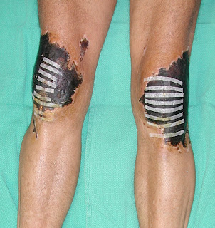
Frostbite after Postoperative Cryotherapy-- Wound Management & Limb Salvage


Picture #1: Skin Necrosis after Frostbite from Cryotherapy
Picture #2: After Debridement of Soft Tissues, use of Negative Pressure Dressing
Picture #3: Bilateral Free Flap (Rectus Muscles + Skin Graft) on both Knee Wounds
Picture #4: Bilateral Limb Salvage, Patient is Weight Bearing and Walking

Severe Frostbite of the Knees after Cryotherapy (Excerpts from original article by Lee CK, Pardun J, et al, Orthopedics 30(1):63-4, 2007 Jan)

"Abstract:

We present a case report of a patient who sustained full thickness soft tissue injuries over the bilateral knees after patellar tendon repairs and postoperative cryotherapy. The injury was severe enough to require bilateral microvascular free tissue transplants to cover both knee surfaces. Complications from cryotherapy have been reported in the literature, but are not common; this represents an extreme example. We review the literature and discuss treatment and prevention protocols.

Cryotherapy has been used to treat pain and inflammation since the
time of Hippocrates.1 Ice, snow,cold water, and cold compresses have been used to treat a multitude of soft-tissue traumas.2 More recently, cryotherapy
has been used increasingly in sports injuries and in the postoperative orthopedic setting.3 However, there have been a number of reports of complications from cryotherapy– most commonly frostbite and peripheral nerve injury–that point out itsbenefi ts but also its dangers.1-5 These previously reported complications has been diverse in location and severity. This article reports a significant complication of cryotherapy as a result of a relatively common regimen of application of ice packs to knees in a postoperative setting...

This case represents a severe frostbite injury after cryotherapy. With proper instruction and use of the cooling device, these complications are mainly avoidable. The patient had minimal padding between the cooling wrap and skin. In addition the patient used the device continuously for two weeks. It was likely that thermal injury occurred the moment the dressing was applied until the patient first took off the dressing two weeks later.

Frostbite occurs by the formation of ice crystals in the intracellular and extracellular space. During the cooling process, the extracellular ice crystals form and osmotic pressure increases, drawing water out of the cells. This leads to intracellular dehydration with an increase in intracellular electrolytes, proteins and enzymes which lead to cell death. Additionally, there is vascular endothelial damage leading to intravascular thrombosis and reduced blood flow. AV shunting occurs at the capillary level and end organ tissue damage is compounded. During the warming process, there is an influx of fluid back into the cells causing intracellular swelling. The warming process also allows reflow, vasodilation and reactive hyperemia to occur leading to increased inflammatory mediators, causing further cell death.

Cryotherapy works by three main processes. First is the reduction in the inflammatory process by inducing a hypometabolic state. Decreasing inflammation decreases the amount of cellular damage by inflammatory mediators, ultimately reducing the amount of capillary permeability and thereby decreasing edema. Second is the decrease in hematoma formation which is produced from vaso-capillary constriction and decreased blood flow. Finally is the induction of analgesia by cold. This is thought to be due to decreased nerve conduction velocity and decreased muscle spasm. In combination, cryotherapy is an ideal postoperative therapy which decreases pain, inflammation, hematoma, and the amount of postoperative narcotic usage...

Current recommendations for cryotherapy include 20-30 minutes of cryotherapy with a maximum time of 40 minutes, always with a protective covering (usually a towel) between the cryotherapy wrap and the skin. The cycle can be repeated every 2 hours while the patient is awake....

Although the incidence of complications from cryotherapy appear rare, estimated at 0.00225%,5 it is likely that this number is an underestimation as there are many unreported cases in conjunction with the increased use of continuous cryotherapy in the postoperative setting. We see this as an opportunity to emphasize the importance of education between the patient and doctor about this device and point out its potentially devastating risks.

Friday, May 29, 2009

Outpatient Wound Care Center in San Francisco 2009


San Francisco Wound Care & Reconstructive Surgery Center


We are proud to annouce the opening of an outpatient wound care center in San Francisco on June 1, 2009. It will consist of a multidisciplinary team of reconstructive plastic surgeons, orthopedic surgeons, general and vascular surgeons, podiatrists, endocrinologists and internal medicine, and dedicated wound nurse practitioners. Our team is composed of surgeons and physicians from the University of California, San Francisco (UCSF) as well as the community at St. Mary's Medical Center.

We will focus on all wound types, both acute and chronic, and of any complexity. These wounds can be from traumatic, vascular, infectious, metabolic, and cancer and radiation. We will have every tool and cutting edge option available to heal the simple to most difficult wounds.


Our office is at 450 Stanyan (Main Building of St.Mary's), 2nd Floor at the PROS Center. Telephone number is 415 750 5588. Email: sfwounds@gmail.com Please ask to speak to Josie Gomez, our wound nurse practioner.